The Current State of Lung Imaging
Diagnostic lung imaging has evolved significantly, from the earliest lung volume testing in the 1800s, rectilinear scanning in the mid-1960s, the first commercially available CT (computerized tomography) scanner in the 1970s,1 and in 1977, the first human MRI scan,2 allowing for higher resolution soft-tissue imaging than ever before.
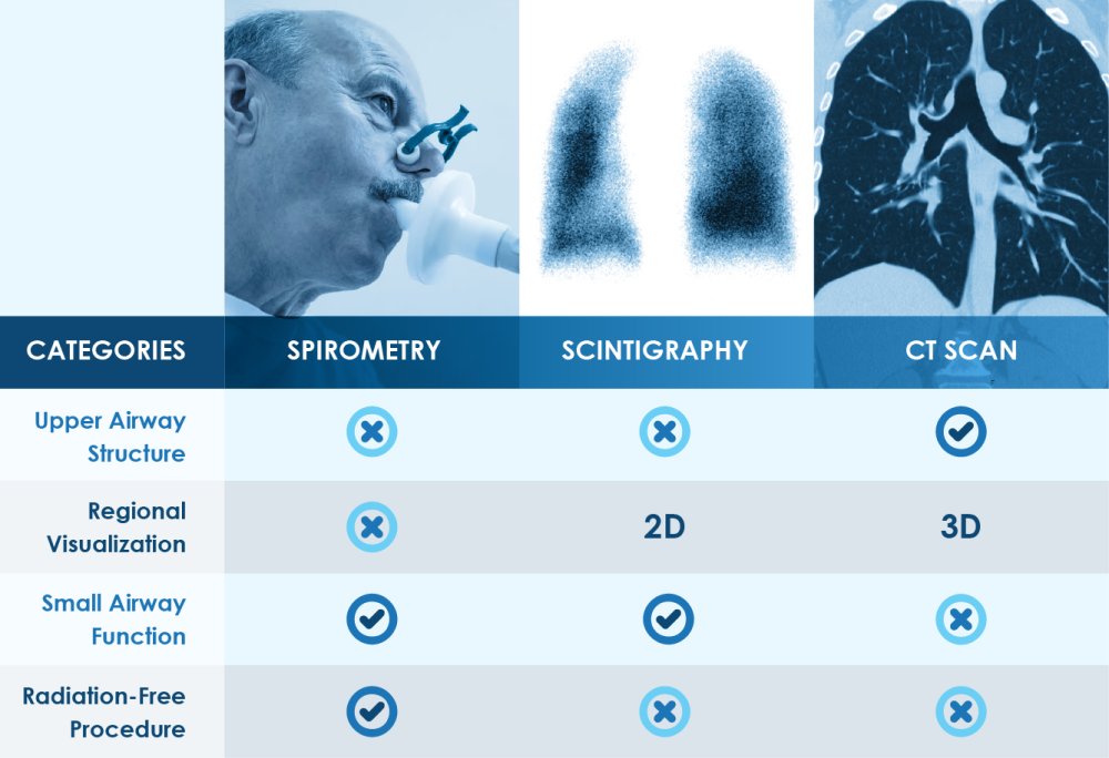
Despite many advancements, the lungs are one of the most complex organs for functional imaging. Most lung diagnostic and imaging modalities currently in use show some of the upper airway structure, but with varying degrees of detail and accuracy. Current diagnostic tools, such as scintigraphy and CT Scan, carry their own inherent limitations and risks, making repeated diagnostic and therapeutic use prohibitive to both the patient and the test administrator.
Current lung diagnostic and imaging modalities can’t visualize the functional network of over 300 million alveoli, which is needed to increase our understanding of how the lungs are working and how effectively ventilation is taking place regionally.
See How Xenon MRI Works
Watch how hyperpolarized xenon-129 gas is inhaled through a single 10-to-15-second breath-hold and distributed to image the smallest airways.
Current Lung Diagnostic Methods Have Advantages and Limitations
There are a number of lung diagnostic and imaging modalities in use today with various advantages and limitations:
Radiation Exposure Risks Complicate Lung Imaging

Cumulative radiation exposure makes longitudinal imaging problematic.
mSv (millisievert) per procedure
Xenon MRI for Lung Imaging
References & Other Information:
1. Beckmann EC. “CT scanning the early days”. Br J Radiol. 2006;79(937):5–8.doi:10.1259/bjr/29444122.
2. Wakefield J. “The “Indomitable” MRI”. Smithsonian Institution. 1 June 2000, https://www.smithsonianmag.com/science-nature/the-indomitable-mri-29126670/.